Visibility plays a crucial role in the successful transfer of technology and the adoption of new technologies in their go-to-market journey. When a new technology is highly visible, it attracts attention from potential adopters, investors, and stakeholders. This visibility can lead to increased interest and awareness, which are essential for fostering acceptance and integration.
This page presents our clients’ latest innovations: smart, scalable solutions designed to meet real-world challenges with elegance and precision. From early-stage prototypes to market-ready systems, these technologies reflect a new generation of inventive thinking and strategic execution.
Technology transfer is essential for driving progress and addressing challenges in various sectors. By fostering collaboration and the exchange of knowledge, we can unlock the full potential of new technologies and create a brighter, more sustainable future. What you see here is more than tech—it’s a glimpse into the future we’re building, one invention at a time
We are thrilled to introduce these ground-breaking technologies that are poised to revolutionize various industries and are now seeking adopters, commercial partners, and licensees.
We have been or still are actively seeking commercial partners / licensees to bring these inventions to the market.
We believe that together, we can make a fantastic impact!

Cardiolipin-specific fluorescent probe
2025 – Cardiolipin is a unique phospholipid that plays a crucial role in the function of mitochondria, the powerhouse of the cell. It is predominantly found in the inner mitochondrial membrane, where it helps maintain the structural integrity and optimal function of various mitochondrial proteins and enzymes. Cardiolipin is essential for the proper functioning of the electron transport chain, which is responsible for producing ATP, the primary energy currency of the cell. Additionally, cardiolipin is involved in the regulation of mitochondrial dynamics, including processes such as fusion, fission, and apoptosis. Due to its critical role in mitochondrial function, cardiolipin has been the subject of extensive research, particularly in the context of mitochondrial diseases and metabolic disorders.
Fluorescent probes are powerful tools used in biological research to study various cellular and molecular processes. These probes are molecules that emit light when excited by an external energy source, such as a laser or UV light. Fluorescent probes can be designed to target specific biomolecules, such as proteins, nucleic acids, or lipids, allowing researchers to visualize and track these molecules in real-time. They are widely used in techniques such as fluorescence microscopy, flow cytometry, and fluorescence in situ hybridization (FISH). The versatility and sensitivity of fluorescent probes make them invaluable for studying complex biological systems, including cell signaling, protein-protein interactions, and gene expression. By providing detailed insights into the spatial and temporal dynamics of cellular processes, fluorescent probes have significantly advanced our understanding of biology and medicine.
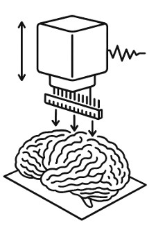
Imaging of biological tissues.
2025 – This patented innovation1 enables the high-resolution electrical characterisation of the features of biological samples in two dimensions (2Dmap), with minimal sample preparation, under mild water-based conditions.
The core idea is simple yet powerful: It becomes possible to obtain a spatial map of tissue permittivity and conductivity under a liquid water-like mediator, meaning the biological material can remain almost untreated and close to its natural state.